Post on Thornwaldt’s Cyst

89e365f443439166f8c9210754abc6_big_gallery
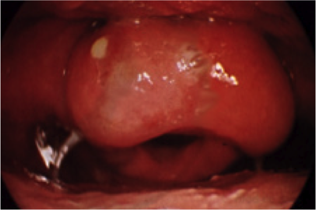
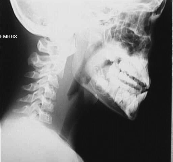
A Thornwaldt cyst (also spelled as a Tornwaldt cyst or Thornwald cyst) is a common incidental benign midline nasopharyngeal mucosal cyst.

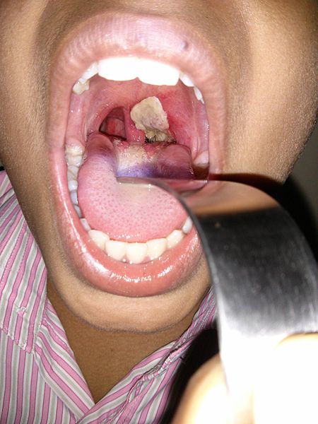
Thornwaldt’s cysts are classified as crusting and cystic 8. They form as a result of retraction of the notochord where it contacts with the endoderm of the primitive pharynx.
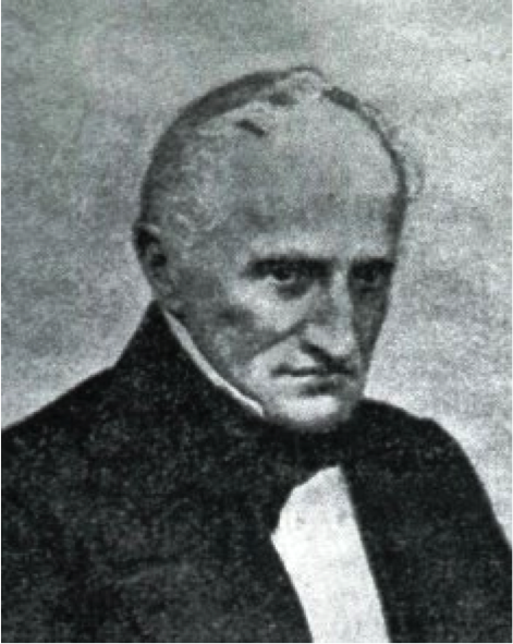
This is believed to happen at about the 10th week of embryonic development. Closure at the orifice results in so called cystic type, while crusts adhering to the orifice without closing result in crust type 8. The cyst is lined by respiratory epithelium and accumulates with fluid with variable proteinaceous content, inflammation can occur due to obstruction.It is named after Gustav Ludwig Thornwaldt, German physician, 1843-1910

Thornwaldt’s Cyst

Post on Choanal Atresia

choanal_atresia2_mediumChoanal Atresia is a rare malformation of the posterior nasal cavity. It results from persistence of the bucconasal membrane of Hochstetter and affects about 1 in 6,000 live births. This can be a frustrating and frightening encounter as infants are obligate nasal breathers. Females are affected twice as often as males and unilateral is twice as common as bilateral. When the atresia is unilateral, the right side is affected twice as often as the left. 90% of atresias are boney whereas 10% are membranous, about half of the atresia cases are associated with other congenital anomalies such as the CHARGE syndrome.

Choanal Atresia

Post on Nasolacrimal Duct Cyst

180px-Dacryocystocele_nose_view002The etiology of congenital nasolacrimal duct obstruction is most commonly a membranous obstruction at the valve of Hasner at the distal end of the nasolacrimal duct. General stenosis of the duct is the second most common cause of duct obstruction. Congenital proximal lacrimal outflow dysgenesis involves maldevelopment of the punctum and canaliculus. Proximal outflow dysgenesis can occur concurrently with distal obstruction. Congenital lacrimal sac mucocele or dacryocystocele occurs when there is a membranous cyst extending from the distal end of the duct into the nose. The nasolacrimal duct sac is filled at birth with clear amniotic fluid.

0392-100X.28.298.fig1Congenital nasolacrimal duct obstruction occurs in approximately 5% of normal newborn infants. The blockage occurs most commonly at the valve of Hasner at the distal end of the duct. There is no sex predilection and no genetic predisposition. The blockage can be unilateral or bilateral.  The rate of spontaneous resolution is estimated to be 90% within the first year of life. Thus timing intervention with resolution makes this a “winner’s game”.

Nasolacrimal Duct Cyst

Post on Pyriform Apeture Stenosis

inline_192_pyriform_aperture_stenosis_mediumPyriform aperture stenosis (PAS) is a very rare congenital anomaly where the anterior opening of the nose is narrow secondary to overgrowth of the maxillary bone. PAS is sometimes associated with other abnormalities, including the presence of a single central incisor tooth and pituitary abnormalities.
What are the causes of pyriform aperture stenosis?
Pyriform aperture stenosis is present at birth and can be associated with other abnormalities. There is no known specific cause.

Pyriform Aperture Stenosis

Post on Epiglottitis

UntitledAcute Epiglottitis is fortunately a rarity since the regular use of the H. Influenze type B vaccination.  Other nomenclature for the disease include Supraglottitis, Oedema glottis, and Supraglottic Croup.


Epiglottitis

Post on Angioedema

UntitledAngioedema (or Quincke’s edema) is inflammation of the deep dermis or submucosa. It is non pitting, non dependent, non tender, non erythematous, usually assymmetric.  The term angiioneurotic oedema was a misnomer as it was initially thought to be a nervous system disorder. It can be life threatening if it involves the airway. It can be congenital, idiopathic, or drug induced. Ace inhibitor blood pressure medications are common offenders causing this condition. NSAIDS and food allergies are the other big culprit. A thorough allergy history and testing are paramount. Exciting new drugs are on the horizon for this condition.

Angioedema

Post on Retropharyngeal Abscess

UntitledMost commonly seen in infants and young children, retropharyngeal abscess (RPA) is an abscess located in the tissues in the back of the throat behind the posterior pharyngeal wall (the retropharyngeal space). Because RPA’s typically occur in deep tissue, they are difficult to diagnose by physical examination alone. RPA is a relatively uncommon illness, and therefore may not receive early diagnosis in children presenting with stiff neck, malaise, difficulty swallowing, or other symptoms listed below. Early diagnosis is key, while a delay in diagnosis and treatment may lead to death. Parapharyngeal space communicates with retropharyngeal space and an infection of retropharyngeal space can pass down behind the oesophagus into mediastinum. [1] RPA’s can also occur in adults of any age.

Retropharyngeal Abscess


Post on Ludwig’s Angina

UntitledLudwig’s angina, otherwise known as angina ludovici, is a serious, potentially life-threatening cellulitis[1][dead link], or connective tissue infection, of the floor of the mouth, usually occurring in adults with concomitant dental infections and if left untreated, may obstruct the airways, necessitating tracheotomy. It is named after the German physician, Wilhelm Friedrich von Ludwig who first described this condition in 1836.

Ludwig’s Angina

Post on Diptheria

450px-Dirty_white_pseudomembrane_classically_seen_in_diptheria_2013-07-06_11-07Diphtheria (Greek διφθέρα (diphthera) “pair of leather scrolls”) is an upper respiratory tract illness caused by Corynebacterium diphtheriae, a facultative anaerobic, Gram-positive bacterium.[1][2] It is characterized by sore throat, low fever, and an adherent membrane (a pseudomembrane) on the tonsils, pharynx, and/or nasal cavity.[3] A milder form of diphtheria can be restricted to the skin. Less common consequences include myocarditis (about 20% of cases) [4] and peripheral neuropathy (about 10% of cases).[5]

Diptheria

Styloid Process and Eagle’s Syndrome

Gray907The Styloid Process is a pointed piece of bone that extends down from the skull, just below the ear. It serves as an anchor point for ligaments and muscles associated with the tongue and larynx.

The stylohyoid ligament, stylomandibular ligament, styloglossus muscle, stylohyoid muscle and stylopharyngeus muscle are attached to the styloid process.  (3 muscles innervated by 3 cranial nerves attach to the styloid process—–stylohyoid – C.N. VII, stylopharyngeus – C.N. IX, and styloglossus – C.N. XII)

imagesA small percentage of the population suffers from a condition called Eagle Syndrome which causes an elongation of the styloid process and stylohyoid ligament calcification. When someone with Eagle Syndrome swallows, the tissue of the throat rubs against the styloid process, resulting in glossopharyngeal nerve pain. Eagle Syndrome was first described by Watt Weems Eagle, an otorhinolaryngologist, in 1937.