 Most commonly seen in infants and young children, retropharyngeal abscess (RPA) is an abscess located in the tissues in the back of the throat behind the posterior pharyngeal wall (the retropharyngeal space). Because RPA’s typically occur in deep tissue, they are difficult to diagnose by physical examination alone. RPA is a relatively uncommon illness, and therefore may not receive early diagnosis in children presenting with stiff neck, malaise, difficulty swallowing, or other symptoms listed below. Early diagnosis is key, while a delay in diagnosis and treatment may lead to death. Parapharyngeal space communicates with retropharyngeal space and an infection of retropharyngeal space can pass down behind the oesophagus into mediastinum. [1] RPA’s can also occur in adults of any age.
 RPA can lead to airway obstruction or sepsis – both life-threatening emergencies.[2] Fatalities normally occur from patients not receiving treatment immediately and suffocating prior to knowing that anything serious was wrong.
Causes
RPA is usually caused by a bacterial infection originating from the nasopharynx, tonsils, sinuses, adenoids or middle ear. Any Upper Respiratory Infection (URI) can be a cause. RPA can also result from a direct infection due to penetrating injury or a foreign body.
Symptoms
Symptoms may include stiff neck (limited neck mobility or torticollis)[3], some form of palpable neck pain (may be in “front of the neck” or around the Adam’s Apple), malaise, difficulty swallowing, fever, stridor, drooling, croupy cough or enlarged cervical lymph nodes. Any combination of these symptoms should arouse suspicion of RPA.
Treatment
RPA’s frequently require surgical intervention. A tonsillectomy approach is typically used to access/drain the abscess, and the outcome is usually positive. Surgery is done without general anesthesia since there is risk of rupture of abscess during intubation for anesthesia. In complex cases, tracheotomy may be required to prevent upper airway obstruction caused by edema in the neck.
High dose of Intravenous antibiotics are required in order to control the infection and reduce the size of the abscess prior to surgery. Chronic retropharyngeal abscess is usually secondary to tuberculosis and the patient needs to be started on anti tubercular therapy as soon as possible.
Diagnosis
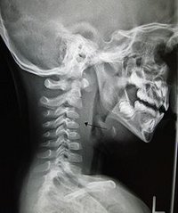
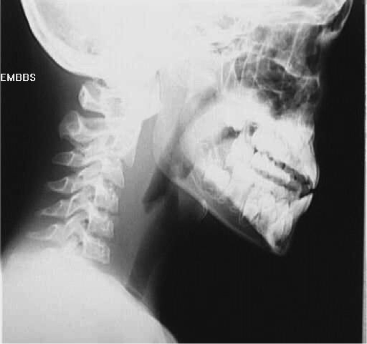
CT with and without contrast is the definitive diagnostic test.
X ray of the neck 80% of the time shows swelling of the retropharyngeal space. If the retropharyngeal space is more than half of the size of the C2 vertebra, it may indicate retropharyngeal abscess.