 Ludwig’s angina, otherwise known as angina ludovici, is a serious, potentially life-threatening cellulitis[1][dead link], or connective tissue infection, of the floor of the mouth, usually occurring in adults with concomitant dental infections and if left untreated, may obstruct the airways, necessitating tracheotomy. It is named after the German physician, Wilhelm Friedrich von Ludwig who first described this condition in 1836.[2][3] Other names include “angina Maligna” and “Morbus Strangularis”. Ludwig’s angina should not be confused with angina pectoris, which is also otherwise commonly known as “angina”. The word “angina” comes from the Greek word ankhon, meaning “strangling”, so in this case, Ludwig’s angina refers to the feeling of strangling, not the feeling of chest pain, though there may be chest pain in Ludwig’s angina if the infection spreads into the retrosternal space. Interestingly, Dr. Ludwig actually died of a neck infection, perhaps even the one that bears his name.
Causes
 Dental infections account for approximately eighty percent of cases of Ludwig’s angina.[4] Mixed infections, due to both aerobes and anaerobes, are of the cellulitis associated with Ludwig’s angina. Typically, these include alpha-hemolytic streptococci, staphylococci and bacteroides groups.[4]
The route of infection in most cases is from infected lower molars or from pericoronitis, which is an infection of the gums surrounding the partially erupted lower (usually third) molars. Although the widespread involvement seen in Ludwig’s usually develops in immunocompromised persons, it can also develop in otherwise healthy individuals. Thus, it is very important to obtain dental consultation for lower-third molars at the first sign of any pain, bleeding from the gums, sensitivity to heat/cold or swelling at the angle of the jaw. Ludwig’s angina is also associated with piercings of the lingual frenulum.[5][6][7]
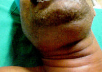
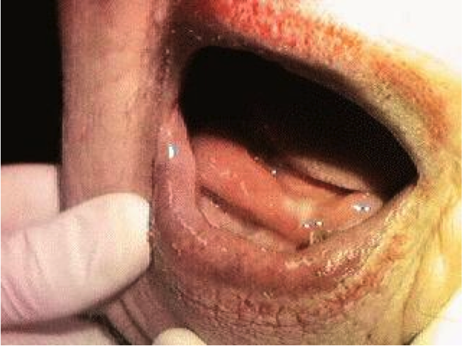
Symptoms and signs
Localisation of infection to the sublingual space is accompanied by swelling of structures in the floor of the mouth as well as the tongue being pushed upwards and backwards.[4]
Spread of infection to submaxillary space is usually accompanied by signs of cellulitis rather than those of an abscess. Submental and submandibular regions are swollen and tender.
Additional symptoms include malaise, fever, dysphagia (difficulty swallowing), odynophagia (pain during swallowing)[4] and, in severe cases, stridor or difficulty breathing. There may also be varying degrees of trismus. Swelling of the submandibular and/or sublingual space is imminent.
Treatment
Treatment involves appropriate antibiotic medications, monitoring and protection of the airway in severe cases, and, where appropriate, urgent maxillo-facial surgery and/or dental consultation to incise and drain the collections.The antibiotic of choice is from Penicillin group.
Incision and drainage of the abscess may be either intraoral or external. An intraoral incision and drainage procedure is indicated if the infection is localized to the sublingual space. External incision and drainage is performed if infection involves the submaxillary space.[4] A nasotracheal tube is sometimes warranted for ventilation if the tissues of the mouth make insertion of an oral airway difficult or impossible. In cases where the patency of the airway is compromised, skilled airway management is mandatory. This entails management of the airway according to the American Society of Anesthesiologists’ Difficult Airway Algorithm and necessitates fiberoptic intubation.