Author Archives: Daniel Todd

Moisturizing Nasal Treatments

Our nose makes over a liter of secretions per day.   To warm, filter and humidify the incoming air in preparation for the lungs is one of the major functions the nose is responsible for.

shoppingBactroban (Mupirocin) ointment is a water miscible ointment that out performs all others in the anterior nose.

shopping-1Pretz Spray Web smallPonaris Nasal Emmolient is also a staple of our practice.  Pretz, Blairex, Nose Better, Nasal Moist, and Rhinaris are all moisturizing sprays.  Entertainer’s Secret is a special formulation used as a throat spray that some have used as a nasal moisturizer. Mayo clinic has compounded rose geranium in sesame oil they say works well.


Holoprosencephaly

photo-7Holoprosencephaly (HPE, once known as arhinencephaly) is a cephalic disorder in which the prosencephalon (the forebrain of the embryo) fails to develop into two hemispheres. Normally, the forebrain is formed and the face begins to develop in the fifth and sixth weeks of human pregnancy. The condition also occurs in other species.

The condition can be mild or severe. According to the National Institute of Neurological Disorders and Stroke (NINDS), “in most cases of holoprosencephaly, the malformations are so severe that babies die before birth.”

When the embryo’s forebrain does not divide to form bilateral cerebral hemispheres (the left and right halves of the brain), it causes defects in the development of the face and in brain structure and function.

In less severe cases, babies are born with normal or near-normal brain development and facial deformities that may affect the eyes, nose, and upper lip.

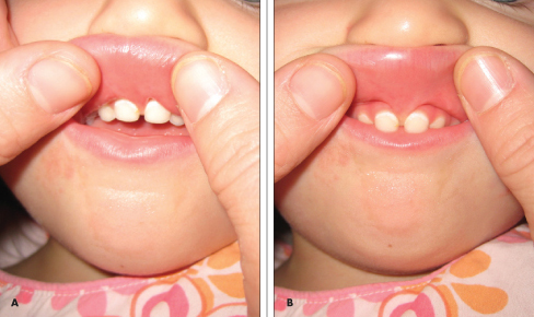
Symptoms of holoprosencephaly range from mild (no facial/organ defects, anosmia, or only a single central incisor) to moderate to severe (cyclopia).

There are three classifications of holoprosencephaly.
Gross pathology specimen from a case of alobar holoprosencephaly.
Alobar holoprosencephaly, the most serious form, in which the brain fails to separate, is usually associated with severe facial anomalies, including lack of a nose and the eyes merged to a single median structure, see Cyclopia
Semilobar holoprosencephaly, in which the brain’s hemispheres have somewhat divided, is an intermediate form of the disease.
Lobar holoprosencephaly, in which there is considerable evidence of separate brain hemispheres, is the least severe form. In some cases of lobar holoprosencephaly, the patient’s brain may be nearly normal.
Syntelencephaly, or middle interhemispheric variant of holoprosencephaly (MIHV), in which the posterior frontal lobe and the parietal lobe are not properly separated, but the rostrobasal forebrain properly separates; it is possible that this is not a variant of HPE at all, but is currently classified as such.[2]
photo-6Holoprosencephaly consists of a spectrum of defects or malformations of the brain and face. At the most severe end of this spectrum are cases involving serious malformations of the brain, malformations so severe that they often cause miscarriage or stillbirth. At the other end of the spectrum are individuals with facial defects which may affect the eyes, nose, and upper lip – and normal or near-normal brain development. Seizures and mental retardation may occur.

The most severe of the facial defects (or anomalies) is cyclopia, an abnormality characterized by the development of a single eye, located in the area normally occupied by the root of the nose, and a missing nose or a nose in the form of a proboscis (a tubular appendage) located above the eye. The condition is also referred to as cyclocephaly or synophthalmia, and is very rare.

Pyriform Aperture Stenosis

Post on Pyriform Aperture Stenosis

inline_192_pyriform_aperture_stenosis_mediumPyriform aperture stenosis (PAS) is a very rare congenital anomaly where the anterior opening of the nose is narrow secondary to overgrowth of the maxillary bone. PAS is sometimes associated with other abnormalities, including the presence of a single central incisor tooth and pituitary abnormalities.
What are the causes of pyriform aperture stenosis?
Pyriform aperture stenosis is present at birth and can be associated with other abnormalities. There is no known specific cause.

Pyriform Aperture Stenosis

Nasal Moisturizing

Our nose makes over a liter of secretions per day.   To warm, filter and humidify the incoming air in preparation for the lungs is one of the major functions the nose is responsible for.

shoppingBactroban (Mupirocin) ointment is a water miscible ointment that out performs all others in the anterior nose.

shopping-1Pretz Spray Web smallPonaris Nasal Emmolient is also a staple of our practice.  Pretz, Blairex, Nose Better, Nasal Moist, and Rhinaris are all moisturizing sprays.  Entertainer’s Secret is a special formulation used as a throat spray that some have used as a nasal moisturizer. Mayo clinic has compounded rose geranium in sesame oil they say works well.


Inverted “V” Deformity

images-5Among the more common complications that occur from a rhinoplasty are asymmetry, difficulty breathing, incompetent internal nasal valves (inverted V deformity) and a pinched tip. Modern rhinoplasty techniques involve far more subtle maneuvers than old-fashioned cutting out cartilage and breaking bones. Since the shape of the nasal tip is determined both by skin thickness and the underlying shape of the nasal cartilages, it used to be taught that to avoid a pinched nasal tip, all the surgeon has to do is avoid cutting out too much cartilage. Otherwise, the nostrils will loose the supportive function of the cartridges and collapse upon themselves thus leading to a “pinch tip” appearance. In actuality, the situation is more complicated than that.

images-6While it is indeed important to leave enough catilage as support, modern techniques of rhinoplasty involve delicately placing precise sutures in order to control the tip and projection of the nasal tip. If the knots are tied ever so slightly tighter than normal, the supportive nature of the cartilage may be overcome by the scar tissue that will develop in the postoperative period. The cartilages will then become concave in appearance thus leading to a pinched tip as seen in the accompanying photo.

Human Bite Injuries

photo-5Eikenella corrodens is a fastidious Gram-negative facultative anaerobic bacillus. It was first identified by M. Eiken in 1958, who called it Bacteroides corrodens.

E. corrodens is a pleomorphic bacillus that sometimes appears coccobacillary and typically creates a depression (or “pit”) in the agar on which it is growing. It grows in aerobic and anaerobic conditions, but requires an atmosphere enhanced by 3–10% carbon dioxide. The colonies are small and greyish, they produce a greenish discoloration of the underlying agar, and smell faintly of bleach (hypochlorite). Only half produce the pitting of the agar considered characteristic. They are oxidase-positive, catalase-negative, urease-negative, and indole-negative, and reduce nitrate to nitrite.

Medical importance
E. corrodens is a commensal of the human mouth and upper respiratory tract. It is an unusual cause of infection and when it is cultured, it is most usually found mixed with other organisms. Infections most commonly occur in patients with cancers of the head and neck,[2] but it is also common in human bite infections, especially “reverse bite” or “fight bite”, or “clenched fist injuries”.[3] It also causes infections in insulin-dependent diabetics and intravenous drug users who lick their needles (“needle-licker’s osteomyelitis”).[4] It is one of the HACEK group of infections which are a cause of culture-negative endocarditis.

The most common pathogens in dog bites are Pasteurella spp. (both Pasteurella multocida and Pasteurella canis)

E. corrodens infections are typically indolent (the infection does not become clinically evident until a week or more after the injury). They also mimic anaerobic infection in being extremely foul-smelling.

Treatment
E. corrodens can be treated with penicillins, cephalosporins, or tetracyclines. It is innately resistant to macrolides (e.g., erythromycin), clindamycin, and metronidazole). It is susceptible to fluoroquinolones (e.g., ciprofloxacin) in vitro, but no clinical evidence is available to advocate their use in these infections.

In popular culture
E. corrodens is mentioned in The Tennis Partner, a memoir by Abraham Verghese.[5]

It is also mentioned in Episode 22, “Punch line” of the sixth season of the television docudrama, “Forensic Files.”

It is also mentioned in Season 3, Episode 8, “Whac-A-Mole” (at 20:51 min) of the TV show House, M.D.

Rhinophyma

unnamed[1]unnamed[1]The term rhinophyma is derived from the Greek rhis (‘nose’) and phyma (‘growth’). Rhinophyma is a large, bulbous, ruddy nose caused by granulomatous infiltration, commonly due to untreated rosacea.  It has often been referred to as “potatoe nose”.  It is most common in white men with a mean age of 50.

Signs and symptoms
rhinophyma_beforeafter_small (1)rhinophyma_beforeafter_smallRhinophyma is characterized by prominent pores and a fibrous thickening of the nose, sometimes with papules.  It is caused by chronic overgrowth of the sebaceous glands and overgrowth of the colonizing bacteria Dermodex Folliculorum. It is associated with the common skin condition rosacea and can be thought of as the end stage of acne rosecea. It can carry a strong psychological impact due to its effect on one’s personal appearance.

Causes
rhinophyma_beforeafter_small (5)rhinophyma_beforeafter_small (4)Alcoholism is mistakenly attributed as a cause of this disease, but heavy alcohol consumption does aggravate the condition due its to chronic vasodilation effects. Rhinophyma may be diagnosed without testing, but a skin biopsy can confirm the diagnosis. Surgical treatment may be beneficial.

Rhinophyma is a slowly progressive condition due to hypertrophy of the sebaceous glands of the tip of the nose often seen in cases of long-standing acne rosacea; it is not a neoplasm. It presents as a pink, lobulated mass over the nose with superficial vascular dilation; it mostly affects men past middle age. Patients seek advice because of the perceived unsightly appearance of the enlargement, or obstruction in breathing and vision.

Treatment
640px-Domenico_ghirlandaio,_ritratto_di_nonno_con_nipote[1]Treating with topical Metrogel seems like a good idea in theory with its activity against Dermodex folliculorum, however, it really does very little in reality. Treatment consists of paring down the bulk of the tissue with a sharp instrument or carbon dioxide laser and allowing the area to re-epithelialise. Sometimes, the tissue is completely excised and the raw area skin-grafted.  I personally have never tried putting a skin graft on this and many of my colleagues think this would be a bad idea.  With the deep and ever present epithelial elements I would expect it to re epithelialize completely in less than a month with just conservative cares (H2O2 and bactroban applied TID).  The painting on the right is from Domenico Ghirlandaio (1449 – 11 January 1494), he was an Italian Renaissance painter from Florence.


Xolair

xolair_largeOmalizumab (trade name Xolair, Roche/Genentech and Novartis) is a humanized antibody originally designed to reduce sensitivity to inhaled or ingested allergens, especially in the control of moderate to severe allergic asthma, which does not respond to high doses of corticosteroids. It has been approved for treating adult and adolescent patients 12 years and older with severe or moderate to severe allergic asthma in more than 90 countries, since its first of such approval in 2002 in Australia. Omalizumab was approved in March 2014 in the European Union and the U.S.A. and in about 10 other countries for treating patients 12 years and above with chronic spontaneous urticaria (CSU) (also referred to as chronic idiopathic urticaria or CIU), which cannot be treated with H1-antihistamines. CSU is not an allergic disease. Presently, the drug is being actively studied in clinical trials for various allergic diseases and some non-allergic diseases, especially skin diseases.

Omalizumab is a recombinant DNA-derived humanized IgG1k monoclonal antibody that specifically binds to free human immunoglobulin E (IgE) in the blood and interstitial fluid and to membrane-bound form of IgE (mIgE) on the surface of mIgE-expressing B lymphocytes.[1] Unlike an ordinary anti-IgE antibody, it does not bind to IgE that is already bound by the high affinity IgE receptor (FcεRI) on the surface of mast cells, basophils, and antigen-presenting dendritic cells.[2]

IgE is commonly involved in type I hypersensitivity, which manifests the most prevalent allergic diseases. It has been estimated that as high as 20 to 40% of the populations who live a western lifestyle in economically advanced countries are affected by allergy and seek medical help.[3] In the U.S., 8% of adults and 10% of children have asthma.[4] Allergy occurs more frequently in individuals with higher serum IgE levels, though some allergic individuals have very low serum IgE, and some people with very high IgE have no allergic problems.[2]

Labial Frenulectomy

Frenum_composite 1106CFP_PC_Frenum_AB-1A labial frenectomy is a form of frenectomy performed on the lip.

The labial frenulum often attaches to the center of the upper lip and between the upper two front teeth. This can cause a large gap and gum recession by pulling the gums off the bone. A labial frenectomy removes the labial frenulum. Orthodontic patients often have this procedure done to assist with closing a front tooth gap. When a denture patient’s lips move, the frenulum pulls and loosens the denture which can be uncomfortable. This surgery is often done to help dentures fit better.

The removal of the frenulum does not cause any adverse effects to the lip and mouth.  The real question is if it has any beneficial effects.

“Potential” benefits include better feeding, diminished decay of the front incisors, and avoiding a central diastema (gap in the front teeth).  I remain a bit skeptical that the benefits exist.

Congenital Cholesteatoma

Congenital+cholesteatomaKeratin-filled cysts that grow medial to the tympanic membrane are considered to be congenital if they fulfill the following criteria: mass medial to the tympanic membrane,
normal tympanic membrane, no previous history of ear discharge, perforation or ear surgery.
Congenital cholesteatomas occur at three important sites: the middle ear, the Petrous apex, and the cerebropontine angle. They are most often found deep to the anterior aspect of the ear drum, and a vestigial structure, the epidermoid formation, from which congenital cholesteatoma may originate, has been identified in this area.