Author Archives: Daniel Todd

Middle Ear Treatments

There was an interesting study on the middle ear volume, and it ended up being about 1.4 ml in non diseased individuals.  In reality, you rarely get a full ml of fluid into the middle ear when you inject it.  There is a number of reasons we put medications directly into the middle ear, such as Meneir’s Disease, sudden sensorineural hearing loss, etc…  We are typically using an anti inflammatory such as dexamethasone or an ablative medication such as Gentamycin.  Sometimes we combine the agents.  When I use the Dexamethasone, I do no dilute, and I put in as much as the middle ear will handle.  I typically anesthetize the ear with phenol or EMLA and have patient stay laying in the injection position without swallowing for 20  minutes if possible.


Challenging scalp lesions

Exposed calvarium represents an interesting challenge in reconstructing scalp defects.  In order to lay on a skin graft, tissue such as a temporalis flap needs to be mobilized to give a base for the graft to survive on.  Otherwise, large “pinwheel” type flaps can be constructed to close this primarily with good success.

This defect was closed with a double layer temporalis muscle flap underlying an occipitally based scalp advancement flap.

IMG_1689

Xolair

xolair_largeOmalizumab (trade name Xolair, Roche/Genentech and Novartis) is a humanized antibody originally designed to reduce sensitivity to inhaled or ingested allergens, especially in the control of moderate to severe allergic asthma, which does not respond to high doses of corticosteroids. It has been approved for treating adult and adolescent patients 12 years and older with severe or moderate to severe allergic asthma in more than 90 countries, since its first of such approval in 2002 in Australia. Omalizumab was approved in March 2014 in the European Union and the U.S.A. and in about 10 other countries for treating patients 12 years and above with chronic spontaneous urticaria (CSU) (also referred to as chronic idiopathic urticaria or CIU), which cannot be treated with H1-antihistamines. CSU is not an allergic disease. Presently, the drug is being actively studied in clinical trials for various allergic diseases and some non-allergic diseases, especially skin diseases.

Omalizumab is a recombinant DNA-derived humanized IgG1k monoclonal antibody that specifically binds to free human immunoglobulin E (IgE) in the blood and interstitial fluid and to membrane-bound form of IgE (mIgE) on the surface of mIgE-expressing B lymphocytes.  Unlike an ordinary anti-IgE antibody, it does not bind to IgE that is already bound by the high affinity IgE receptor (FcεRI) on the surface of mast cells, basophils, and antigen-presenting dendritic cells.

IgE is commonly involved in type I hypersensitivity, which manifests the most prevalent allergic diseases. It has been estimated that as high as 20 to 40% of the populations who live a western lifestyle in economically advanced countries are affected by allergy and seek medical help.  In the U.S., 8% of adults and 10% of children have asthma.  Allergy occurs more frequently in individuals with higher serum IgE levels, though some allergic individuals have very low serum IgE, and some people with very high IgE have no allergic problems.

Pyriform Aperture Stenosis

inline_192_pyriform_aperture_stenosis_mediumPyriform aperture stenosis (PAS) is a very rare congenital anomaly where the anterior opening of the nose is narrow secondary to overgrowth of the maxillary bone. PAS is sometimes associated with other abnormalities, including the presence of a single central incisor tooth and pituitary abnormalities.
What are the causes of pyriform aperture stenosis?
Pyriform aperture stenosis is present at birth and can be associated with other abnormalities. There is no known specific cause.

Merkel-Cell Carcinoma

MERKEL CELL CARCINOMA (TOKER 1972-ENDOCRINE CA OF THE SKIN)

IMG_1692Is caused by the Merkel cell polyomavirus (MCV) 80% of the time, discovered at the University of Pittsburgh in 2008.  It is also known as a cutaneous APUDoma, primary neuroendocrine carcinoma of the skin, primary small cell carcinoma of the skin, and trabecular carcinoma of the skin.  It is a tumor of the tactile merkel cell in the stratum basale of the epidermis.  Histologically it looks like small cell lung cancer with dense cohesive sheets of highly mitotic cells with scant cytoplasm.  They are s-100 positive and are undifferentiated cells of neural crest origin.

It is a rare and aggressive neoplasm that behaves and even looks like an aggressive amelanotic melanoma.  55% present in the head and neck, 20% in the periorbital region, and  primarily in the 6th decade of life.  They are flesh colored and more than 50% overall have regional metastasis.  Surgically we treat them like a melanoma.  However, surgery alone is rarely the treatment plan as they seem to be radiosensitive.  As up to 20% may have distant metastasis at presentation, PET scanning seems appropriate.

In the past we used to say 3 cm margins with lymphoscintigraphy alone for stage 1, 40 GY radiation for stage 2, and induction chemo for stage 3.  Probably palliative chemo for stage 4.

RRP and HPV

gardasil_logo_tcm1908-197175There are over 200 known serotypes of Human Papilloma Virus (HPV). About 79 million Americans are currently infected with HPV. About 7% of adults have HPV in their oral cavity. Only about 3,000 to 13,000 patients in the United States have Recurrent Respiratory Papilllomatosis (RRP). Virtually all of them have active HPV in thier oral cavity. These patients are afflicted with HPV 6 and 11, which affects primarily mucous membranes. It is looking more and more like oral to oral transmission trumps oral genital transmission. It seems to be a different animal all together than cervical HPV. Serotypes 16 and 18 are the types associated with carcinogenesis. There currently is no evidence that spouses should be vaccinated and it does not really seem to be easily transmitted between adults.  Cervarix is a bivalent 16 &18 only vaccine.

This disease has become epidemic, however the treatments and the preventative measures are also gaining momentum.  HPV is the whole reason we really did PAP smears.  The viral changes led to cervical uterine dysplasian and even carcinogenesis.  It was even suggested to consider C-section in women with active infections.

The disease is really divided into the more problematic juvenille onset (>20 lifetime surgeries) vs adult onset (<5 lifetime surgeries).  There may be some correlation with sexual behavior in adult onset, however, this is still controversial. There is no role for vaccination or even counseling or testing partners.

images-1
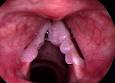
Recurrent Laryngeal RRP may require multiple surgeries.  It seems the best initial response is with the CO2 Laser, however use of the microdebrider (PIPE) may actually give better voice outcomes

Adjuvant therapies such as Acyclovir, MTX, Ribavarin, Mumps vaccine, PPI, Alpha Interferon, Hsp E7, Retinoids, and Intralesional Cidofovir have been touted, but lack strong prospective efficacy studies.  Typically I use the CO2 laser.  Have patients eat green leafy vegetables (Indole-3-carbinols) modulates estrogen metabolism and 1/3 of patients respond.  Inject about 75 mg of Cidofovir in 1 cc after the resection.  I may consider placing on Celebrex (Cox 2 inhibitor) which modulates the over expression of epidermal growth factor by inhibiting cyclooxygenase-2 and prostaglandin E2.   I also may consider injecting Avastin (Bevacizumab) recombinant monoclonal antibody against vascular endothelial growth factor.

Lip Tie

Labial Frenulectomy

Frenum_composite 1106CFP_PC_Frenum_AB-1A labial frenectomy is a form of frenectomy performed on the lip.

The labial frenulum often attaches to the center of the upper lip and between the upper two front teeth. This can cause a large gap and gum recession by pulling the gums off the bone. A labial frenectomy removes the labial frenulum. Orthodontic patients often have this procedure done to assist with closing a front tooth gap. When a denture patient’s lips move, the frenulum pulls and loosens the denture which can be uncomfortable. This surgery is often done to help dentures fit better.

The removal of the frenulum does not cause any adverse effects to the lip and mouth.  The real question is if it has any beneficial effects.

“Potential” benefits include better feeding, diminished decay of the front incisors, and avoiding a central diastema (gap in the front teeth).  I remain a bit skeptical that the benefits exist.

Inverted “V” Deformity

images-5Among the more common complications that occur from a rhinoplasty are asymmetry, difficulty breathing, incompetent internal nasal valves (inverted V deformity) and a pinched tip. Modern rhinoplasty techniques involve far more subtle maneuvers than old-fashioned cutting out cartilage and breaking bones. Since the shape of the nasal tip is determined both by skin thickness and the underlying shape of the nasal cartilages, it used to be taught that to avoid a pinched nasal tip, all the surgeon has to do is avoid cutting out too much cartilage. Otherwise, the nostrils will loose the supportive function of the cartridges and collapse upon themselves thus leading to a “pinch tip” appearance. In actuality, the situation is more complicated than that.

images-6While it is indeed important to leave enough catilage as support, modern techniques of rhinoplasty involve delicately placing precise sutures in order to control the tip and projection of the nasal tip. If the knots are tied ever so slightly tighter than normal, the supportive nature of the cartilage may be overcome by the scar tissue that will develop in the postoperative period. The cartilages will then become concave in appearance thus leading to a pinched tip as seen in the accompanying photo.

Congenital Cholesteatoma

Congenital+cholesteatomaKeratin-filled cysts that grow medial to the tympanic membrane are considered to be congenital if they fulfill the following criteria: mass medial to the tympanic membrane,
normal tympanic membrane, no previous history of ear discharge, perforation or ear surgery.
Congenital cholesteatomas occur at three important sites: the middle ear, the Petrous apex, and the cerebropontine angle. They are most often found deep to the anterior aspect of the ear drum, and a vestigial structure, the epidermoid formation, from which congenital cholesteatoma may originate, has been identified in this area.