Daniel W. Todd, M.D., Midwest Ear Nose & Throat Associates, Sioux Falls, S.D. 57108

 Choanal Atresia is a rare malformation of the posterior nasal cavity. It results from persistence of the bucconasal membrane of Hochstetter and affects about 1 in 6,000 live births. This can be a frustrating and frightening encounter as infants are obligate nasal breathers. Females are affected twice as often as males and unilateral is twice as common as bilateral. When the atresia is unilateral, the right side is affected twice as often as the left. 90% of atresias are boney whereas 10% are membranous, about half of the atresia cases are associated with other congenital anomalies such as the CHARGE syndrome.
Over the past year and a half patients have presented under variable circumstances with choanal atresia. Recent endoscopic techniques and technology, not previously available in the region, help in delivering safe and effective therapy to these infants here in South Dakota.
CASE 1
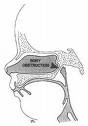
Baby Girl (LT) was born in Clear Lake, SD after an uneventful pregnancy. Her gestational age was 37 ½ weeks and she weighed 2.67 kg. Her APGAR scores were 6 at 1 minute and 7 at 5 minutes. She had copious oral secretions but seemed to pink up nicely when stimulated to cry with oral suctioning. Over the next hour however, she seemed to decompensate and subsequently required oral endotracheal intubation. She was subsequently airlifted to Sioux Falls. Bilateral choanal atresia was diagnosed and ENT was consulted (figure 1).
The malformation was felt to be favorable to an endoscopic repair. This was performed with the pediatric sinus endoscopes, a specially designed sphenoid punch and straight pediatric back-biter. Mucosal sparring techniques were utilized and the microdebrider with pediatric blades was used to finish the technique. Mitomycin C was applied topically to help prevent scarring and a bit of dissolvable sinus packing was left. After 5 more days of remaining on the ventilator the child was returned to the OR and the repair was inspected. Everything looked excellent and topical Mitomycin C was reapplied. The child was subsequently extubated and feeding was started.
CHARGE Syndrome and other associated abnormalities were discovered and the child required a number of other tests and procedures during her stay. Follow up was arranged but the appointments were not kept. Last we heard the child was doing well.
CASE 2
A 5 month old male (DG) who had undergone 2 transnasal repairs with stenting elsewhere presented to our clinic with increasing nasal airway obstruction and difficulty feeding. The child had a high arched palate and boney narrowing of the posterior nasal cavity(Figure 2). He subsequently underwent revision repair with the pediatric endoscopes and required stenting with neonatal argyl chest tubes for 6 weeks. Mitomycin C was applied topically as well. The child did well for 4 months but then required a repeat procedure, again with stenting. He did well again for several months. The family moved out of state and last I heard was again experiencing increasing nasal airway obstruction.
CASE 3
A baby girl (MD) was born term at 6 pounds 11 oz with seemingly no abnormalities. She began showing signs of congestion and had moderate difficulty feeding. Unilateral choanal atresia was diagnosed (figure 3). Conservative cares, such as nasal saline and suctioning, were instituted and the child was discharged. The infant continued to have more and more difficulties and returned to the E.R. after one day. The case was carefully reviewed and in addition to left choanal atresia, right choanal stenosis was diagnosed.
The patient underwent transnasal endoscopic repair. Extreme care was taken not to traumatize any mucosa around the right choana. The right side was enlarged by careful resection of the posterior boney septum (vomer). The pediatric micordebrider was utilized to carefully shape the left posterior choana with maximal mucosal sparring. Mitomycin C was applied topically. The child was extubated the following morning and feeding was successfully instituted. She was discharged the following day. So far the child has done well.
Discussion
The variable severity of choanal atresia and stenosis can make the problem either a medical emergency or a chronically frustrating problem. Initial difficulty with feeding is often the alerting event. As the child tries to feed, there is progressive obstruction and choking due to the aspiration of the milk. This can mimic the presentation of a tracheoesophageal fistula.
Diagnosis is made by the failure to pass cathoders from the nose to the oral cavity. Confirmation and analysis of the extent of disease is further established by thin cut axial CAT scan. Coronal and 3-D reconstruction of the images can be helpful in surgical planning.
Initial management must focus on maintaining the airway. Insertion of an orogastic tube or intraoral (McGovern) nipple can temporarily break the obstructive seal between the tongue and the palate in infants. This in essence allows obligate nasal breathing infants to breathe through their mouth. In reality these infants with bilateral choanal atresia end up being intubated and fed through an orogastric tube. Tracheostomy is rarely indicated.
Once the diagnosis is made and the patient stabilized, a search for associated abnormalities and syndromes must be considered. Infants with bilateral atresia, have more than a 50% chance of suffering other abnormalities. Next, a decision of when and how to fix the malformation must be made. With the newer techniques and endoscopic technologies, earlier intervention is more successful. However, if the child is relatively asymptomatic with only a unilateral problem, waiting for the child to grow is advocated. The literature has clearly shown that multiple procedures are often necessary. An old surgical axiom states: “if you want to close a hole, try to keep it open; if you want to keep a hole open, try to close it.” Unfortunately this has often proven a reality, especially in neonates with very compact anatomy.
Surgical correction is really divided into transnasal or transpalatal approaches. If the infant has multiple nasal and nasopharyngeal abnormalities, such as a highly arched boney palate, then the more invasive transpalatal approach is appropriate. This essentially involves operating through the child’s mouth, raising a palatal flap and drilling out the bone of the post hard palate and choanae. The endoscopic transnasal approach is less invasive and allows for a quicker recovery. It is advocated whenever possible. The question of whether to stent or not must always be raised, but if the opening is made large enough with minimal collateral damage then it is best avoided. Topical mitomycin C, a topical antimetabolite, has proven beneficial in many ear, nose, and throat surgeries to prevent cicatricial scarification and post operative inflammation.
Conclusion
Choanal atresia is a relatively rare malformation of the posterior nasal cavity. It can present with frightening airway obstruction. Newer technologies and endoscopic techniques can allow for earlier, less invasive, and more effective intervention right here in South Dakota.
Bibliography
1) Bluestone CD and Stool SE, editors. Pediatric Otolaryngology. Philadelphia, PA: WB Saunders Company; Chapter 36, 1990.
2) Friedman NR, Mitchell RB, Bailey CM: Management and outcome and of choanal atresia correction. Int J Pediatr Otorhinolarynol 2000 Jan 30; 52(1): 45-51.
3) Lazar RH, Younis RT: Transnasal repair of choanal atresia using telescopes. Arch Otolaryngol Head and Neck Surg 1995 May; 121(5): 517-20.
4) Hartnick CJ: Topical Mitomycin-C following Laryngotracheal Reconstruction: A randomized,double blind, placebo controlled study, Archives of Otolaryngology-Head and Neck Surgery, 126, 1260-1265 (2001)