There are three forms of oral lichen planus.
- The reticular formis the most common presentation and manifests as white lacy streaks on the mucosa (known as Wickham’s striae) or as smaller papules (small raised area). The lesions tend to be bilateral and are asymptomatic. The lacy streaks may also be seen on other parts of the mouth, including the gingiva (gums), the tongue, palate and lips.The reticular form is the easiest to diagnose. The bullas lesions must be differentiated from pemphigoid, chemical burns traumatic ulcers. When they break, they appear as ulcers and need to be differentiated from squamous cell carcinoma.
- The bullous formpresents as fluid-filled vesicles which project from the surface.The atrophic and erosive forms must be differentiated from lichenoid drug reactions,SLE, pemphigoids and other immunobullous disease, candidiasis, erythema multiforme.
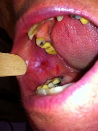
- The erosive forms (Atrophic LP & Ulcerative LP) present with erythematous (red) areas that are ulcerated and uncomfortable. The erosion of the thin epithelium may occur in multiple areas of the mouth (more prominent on the posterior buccal mucosa), or in one area, such as the gums, where they resemble desquamative gingivitis. Wickham’s striae may also be seen near these ulcerated areas. This form may undergo malignant transformation, although this is controversial. The malignant transformation rate is thought to be less than 1%, however it has been reported to be as high as 5%. For any persistent oral lesion of erosive lichen planus that does not respond to topical corticosteroids, a biopsy is recommended to rule out precancerous (premalignant) change or malignant transformation.