 Inflammatory masses are the most common pediatric neck masses encountered. Infectious neck masses may include viral adenitis, bacterial lymphadenitis, atypical mycobacterium, cat-scratch disease, or mononucleosis.
Viral cervical adenitis, the most common type of cervical lymphadenopathy, is self-limited and does not warrant biopsy. It may be a result of adenovirus, rhinovirus, influenza, varicella, herpes or measles. Treatment typically only requires supportive measures. Mononucleosis may present with bilateral posterior cervical adenopathy in the presence of tonsillar enlargement with white exudates. A positive Epstein Barr Virus antibody titer and monospot test is diagnostic and treatment is also supportive with hydration , rest and IV steroids.
Bacterial lymphadenitis, a common indication for hospitalization, is characterized by a recent upper respiratory infection, fever, and tender adenopathy. While the submandibular, retropharyngeal and superior deep cervical nodes are most often involved, bacterial adenitis can affect any lymph node group in the neck. Staphylococcus aureus (80%) and group A beta-hemolytic streptococcus (15%) are the most common pathogens leading to suppuration with a rising incidence of methicillin-resistent staphylococcus aureus in the community. Evaluation of bacterial lymphadenitis involves identifying an erythematous, tender, enlargening neck mass with or without fluctuance. Elevated white blood cell count and C-reactive protein is a common finding but not necessary for diagnosis. Ultrasound is recommended as an initial imaging test to determine size and the presence of fluid (indicating abscess formation). CT imaging, which provides the best resolution for deep neck infections, is advised if there is no clinical improvement after initial treatment, minimal skin changes without obvious fluctuance, or for definitive surgical planning. Needle aspiration can be both diagnostic (culture and sensitivity) and therapeutic if an abscess cavity is present. Management of bacterial neck infections can involve oral antibiotics, IV antibiotics and/or surgical drainage. A trial of oral antibiotics is indicated as an initial treatment when only mild systemic symptoms and skin changes are present. A ten day course of a beta-lactamase resistant antibiotic is recommended. IV antibiotics are indicated in the presence of high fevers, poor PO intake, and marked enlargement of lymph nodes with cellulitis. Phlegmon or minimal suppuration seen on CT scan also predicates the need to initiate IV antibiotic therapy. Double coverage with clindamycin and cefftriaxone is a common regimen. Lack of improvement on IV antibiotic therapy over 48-72 hours or progression in size or fluctuance necessitates CT imaging, if not already done, or immediate surgical incision and drainage. Following surgical drainage, culture driven PO antibiotics are initiated for 10 to 14 days.
Cat scratch disease has an incidence of 9.3/100,000 which presents with cervical adenopathy weeks after inoculation by contact with cats. Children may have fever and malaise and only up to 30% of the nodes may suppurate. Definitive diagnosis is by serology with positive Bartonella hensalae titers. Treatment is primarily supportive but may include a short course of a macrolide antibiotic.
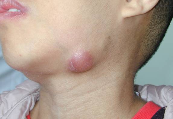
Atypical mycobacterium, the leading cause of cervical tuberculosis in children, affects kids between the ages of 1 and 5. The most common pathogen is Mycobacterium avium-intracellulare and inoculation is from contaminated food, water, or soil. Patients often lack systemic symptoms and present with a < 3 cm lateral submandibular neck mass that may display violaceous skin changes. The mass may spontaneously rupture leading to a draining sinus tract. PPD ought to be placed however a positive result is not needed for diagnosis. Definitive management consists of complete surgical excision and curettage with 1-6 months of antibiotics. A macrolide antibiotic andrifampin are often administered, however type and duration of antibiotic that ought to be used is not clearly defined. There is some thought that atypical mycobacterium infections will resolve on their own regardless of the treatment employed. Nevertheless, it is advised never to perform incision and drainage of the lesion due to a resulting chronically draining sinus tract.